

Do I Need To Repair My Lattice Degeneration
Lattice Degeneration
Vitamins/Supplements What information technology is Where it occurs Progression Symptoms & Causes Treatment Self help Reviews
Lattice degeneration gets its name from the crisscrossing, fine, white lines on the surface of the retina, which an eye physician can run into during examination. Information technology occurs in seven–viii% of the general population, most often in patients with myopia (nearsightedness) and over the age of xx. It as well appears that neither race nor sex is a risk cistron. Approximately 45% of those affected have lattice degeneration in both optics. Patients with lattice degeneration are typically symptom-free.
What is Lattice Degeneration?
Lattice degeneration occurs when the outside edge of the retina (away from the key macula), responsible for peripheral vision, shrinks, thins, develops holes, or otherwise atrophies. Clinical features may include retinal thinning, branching, whitish lines on the retinal surface, and even small holes in the retina. In addition, lesions can class.
Sometimes lesions crusade holes or breaks in the retina, which are atrophic in nature. Atrophic means that they are due to the wasting abroad or diminishing of portions of the retina. Unlike retinal detachment, these breaks and holes are non associated with pulling betwixt the vitreous and retina. They are normally an incidental finding of a dilated ophthalmologic examination that may occur inside the lattice (present in 25–35%) and result from progressive retinal thinning. The fine lines seen in lattice degeneration are present in roughly just x% of all lesions. The other 90% are known as uncomplicated lattice degeneration, with lesions but no tears, holes, or breaks.
Where Does information technology Occur?
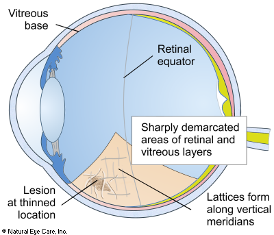
Lattice degeneration occurs between the retinal equator and the vitreous base.
The retinal equator is an imaginary boundary midway between the front and the back of the eyeball. The vitreous base lies toward the front of the middle where the retina, vitreous membrane, and pigmented layers firmly attach to each other. Lattice degeneration may show up equally sharply demarcated areas of thinning. The thinning typically involves the vitreous and the inner layers of the retina. Sometimes thinning also occurs in the retinal pigment layer and rarely along the fine blood capillaries. The lattices usually form along the vertical meridians and perpendicular to the retinal equator.
Progression
Several things happen as the condition develops:
- the retina atrophies, forming a lesion, and becomes thinner due to obstructed blood supply
- larger blood vessels stiffen and become clogged
- the vitreous gel becomes more liquid over the retina lesion
- the vitreous contracts away from the retina and at the same time becomes more strongly attached to the retina, trigger-happy it, detaching it, or causing holes and breaks in information technology
Sometimes retinal thinning is then great that holes develop through the retina at the lesion. The vitreous is then able to movement through the retina into the space behind the retina contributing to retinal detachment. that a total-thickness pigsty atrophies through the retina at the lattice lesion. The overlying liquefied vitreous has the power to pass through the hole into the subretinal space and for 2% of patients, leads to "rhegmatogenous" retinal detachment. In this example the fluid is accumulating between the retina and the pigment layer (RPE).
Symptoms
Patients are ordinarily over 20 and rarely have symptoms, except for possibly noticing flashing lights (photopsia), a sudden onset of eye floaters, or partial loss of peripheral vision. Patients often accept myopia (nearsightedness).
Causes
Heredity is likely a risk factor. A family history and certain variants in the COL4A4 gene may be risk factors. Lattice is inherited in an autosomal ascendant manner; the affected parent having one mutated gene tin can pass it on to a child.
Myopia increases hazard. Lattice is associated with those who accept high myopic eyes, and its prevalence may be associated with increasing axial length, reaching 15% in the longest eyes. This is especially true of axial centre lengths betwixt 29–30mm and 25–27mm.
Poor circulation. Lattice degeneration may also be linked to an bereft blood supply or a claret supply with insufficient nutrients reaching the retina. From the front of the center, to the retinal equator, the retina is supplied with nutrients by the posterior ciliary artery. When its circulation is degraded or obstructed, a "watershed zone" develops where at that place are fewer microcapillaries.
Other conditions. Researchers have also constitute a high rate of lattice degeneration in patients with full-thickness macular holes.
 Come across Vitamins & Supplements to support the retina and overall eye health.
Conventional Treatment
No standardized treatment exists for lattice degeneration. Clinical studies of interventions to prevent retinal disengagement in lattice patients have been evaluated, but reviewers found that controlled trials were lacking, and no conclusions could be drawn. In certain cases where retinal holes or tears are present, a doctor might recommend laser photocoagulation or cryotherapy (extreme common cold light amplification by stimulated emission of radiation therapy), with the goal of sealing the expanse surrounding the tear. What mode is chosen depends on the location of the tear and whether in that location is fluid associated with it.
In most cases of lattice degeneration, no treatment is necessary. Treatment is recommended when a retinal tear occurs, if the other centre has had a retinal detachment, or if at that place is a strong family unit history of retinal detachments. Surgical intervention may exist required if vitreous fluid is institute to be leaking into the retina, which can complicate the lattice and atrophic holes, or if it is found in a patient who has suffered a previous retinal detachment (in an opposite eye).
Preventative or condom treatment is indicated if there are tractional (pulling) tears due to vitreous detachment, or lesions are nowadays in one center when there is a retinal detachment in the other eye.
Self Assist
Since poor circulation and nutrient deficiency have been identified as possible contributing causes for lattice degeneration, there may exist natural ways to amend these situations, thereby boosting retinal wellness. Even in the case where lattice degeneration is inherited, the body is nevertheless trying to maintain healthy vision. So targeted supplements (antioxidants, in particular) and a healthy lifestyle can assistance to cease or slow down the progression of the illness and back up healthy vision. Certain nutrients such as lutein, zeaxanthin,omega-three fatty acids, vitamin D3, and herbs such equally ginkgo or bilberry may be helpful.
Daily juicing of organic vegetables and fruits. Our recipe to support retinal health is a combination of the following: ginger, garlic, parsley, leeks, beets, cabbage, carrots, spinach, celery, apples, grapes, raspberries, lemon, chlorophyll, wheat grasses - (not besides much fruit). See more on juicing.
Lattice Degeneration News
Want to acquire more than? Encounter our weblog news on lattice degeneration.
Related Conditions
- Lattice degeneration occurs more frequently (40% more than) of patients with retinal disengagement.
- Degenerative myopia, which is similar to macular degeneration
- Marfan'southward syndrome - this is a life-threatening disorder of connective tissue which can affect body systems, such as the heart. Harm due to Marfan can manifest every bit retinal detachment, dislocated lenses, nearsightedness, astigmatism, binocular conditions, glaucoma, and cataracts in children. 75% of cases are genetically caused.
- Stickler syndrome - another genetically-based disorder, is characterized by a somewhat flattened face. Stickler is a joint disorder that affects vision, hearing and joint problems. It is besides known as progressive arthro-ophthalmopathy.
- Ehlers-Danlos syndrome - is another status involving collagen that is besides flexible resulting in loose articulation dislocations, delicate skin, poor muscle tone. The eye (especially the cornea and sclera, is comprised of generally connective tissue, and like lattice degeneration, this condition can result in retinal disengagement.

Source: https://www.naturaleyecare.com/eye-conditions/lattice-degeneration/
Posted by: jaredfroneam1996.blogspot.com
0 Response to "Do I Need To Repair My Lattice Degeneration"
Post a Comment